
Dark-field microscopy
For the visualization of cellular structures, in general, it is always necessary to use means that increase the resolution, in view of the invisibility to the naked eye of these structures. Optical microscopy is the most common method of observation, useful in most studies and widely used in clinical analysis laboratories. For further info, click here: dark field microscopy
Through traditional staining techniques, the samples of interest are previously treated, making it possible to identify them with a wealth of details. The dye penetrates cell structures through the membrane's permeability, allowing the light emitted by the microscope to differentiate the sample from the background.
Blood cells are smeared on a glass slide with a small sample of the patient's blood, subsequently treated with a trio of dyes called Panotic. The first reagent is a fixative that will act as a preservative, preserving cellular structures and blocking chemical reactions in progress. The second is an acid dye that will dye the cytoplasm and basic cell structures pink. The third will dye the core purple-blue.
With the help of an optical microscope, the light reflects the sample and the background in question, but only the cellular structures of interest will be stained. This method makes only structural and quantitative observation possible, as the process ends up immobilizing and drying the cells. The shades will depend on the exposure time. Some structures more sensitive to pH may show sudden changes than when observed in vivo.
Each type of sample will need specific treatment. To identify the presence of intestinal parasites, for example, it is necessary to use a solution of Lugol in the diluted feces, as urinary crystals can hardly be colored due to its practically impermeable structure. In these cases, visualization is more difficult, requiring the professional to master the main characteristics of these structures in order to perform the recognition.
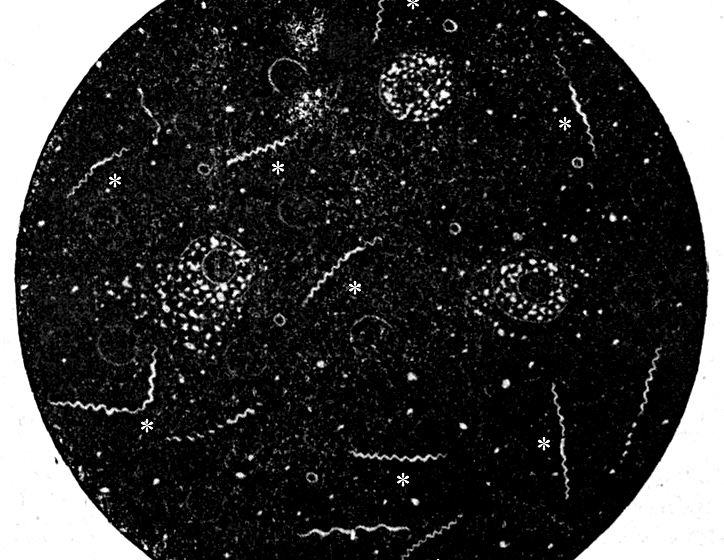
Aiming at a rich identification in details came the Dark Field Microscopy, a technique used in situations in which the samples do not allow an effective staining or in analyzes of live cells, that is, samples that have not undergone immobilization treatment, the traditional fixation.
Structurally, the Dark Field Microscope is very similar to the Common Optical Microscope. Both have Condenser, Eyepiece and Objective Lenses, in addition to the light source at the bottom. The only difference is the presence of an Opaque Disc below the Condenser. While the light emitted by the Common Optical Microscope passes through the entire sample, reflecting the bottom of the area around the object of interest, in Dark Field Microscopy (MCESV) the Opaque Disc will be responsible for preventing the emitted light from passing through the Condenser entirely . In this way, only a small fraction will reach the sample while the majority will deviate. By focusing the beam only on the structures of interest, the observer will not suffer interference from the bottom of the sample, resulting in a more defined visualization.
Despite being useful in the visualization of countless microorganisms such as Spiroquetas Leptospira interrogans, which causes Leptospirosis and Treponema pallidum, responsible for syphilis, Dark Field Microscopy (MCESV) has been widely used in the study of blood cell morphology in living blood. Through the rapid collection of a small amount of blood, still in the office, it is possible to quantitatively and qualitatively evaluate a patient sample, determining the degree of abnormality through parameters such as size, color, type, shape and activity of the cells.

